


Hattrick against brain and spinal cord tumors in children
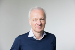
They are rare and occur in both the brain and spinal cord of children: Tumors called ependymomas. Three scientists from Prof. Ulrich Schüller's research group at the Research Institute "Kinderkrebs-Zentrum Hamburg" have now achieved groundbreaking results that will make it possible to better diagnose and treat these tumors in the future.
Ependymomas account for almost five percent of all tumors of the central nervous system in children. The tumor cells arise from ependymal cells, which line the inner walls of the brain's ventricles and the spinal canal. Effective treatments are currently limited to complete surgical removal of the tumor and radiation therapy. In young children in particular, efforts are made to avoid irradiating the brain in order to avoid permanent damage. Knowledge to better classify these tumors and predict their evolution is essential to make treatment as gentle and effective as possible for each patient.
Better assessment of relapse risk
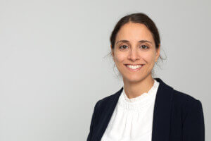
In the case of spinal ependymoma (SP-EPN), there is now more clarity. Dr. Sina Neyazi has studied these tumors, which occur in the spinal cord, and found that there are two subtypes that progress differently. Subtype A is more likely to recur, meaning the tumor will come back, while subtype B is unlikely to recur. The two subtypes differ at the molecular level. Among other things, certain changes in the genetic material, known as NF2 mutations, occur in the more severe subtype A, while these mutations are not found in subtype B, or only rarely.
Prof. Schüller explains how these new findings translate into clinical practice: "Nobody dies from these spinal cord tumors. But what do I tell the parents after the tumor has been surgically removed? Should they come back in six months, a year, or two years to have their child examined? Or should they assume that the tumor has progressed so well that they only need to come back if symptoms recur?”
With Dr. Neyazi's results, it is now possible to classify the course of the disease much more clearly by molecular analysis of the tumor tissue. Detailed insights into the molecular properties of these tumors are also essential for the development of new treatment strategies.
Most comprehensive ependymoma analysis to date
In her work, Lara Pohl examined 2,023 ependymoma data sets. In doing so, she created a data set that provides reliable information on how certain molecular characteristics affect the progression of the tumor and the survival rate.
The last comparable analysis was performed in 2015, but it included only 500 samples, which meant that some tumor classes were still missing. Pohl was now able to fill in the missing data, verify the findings from that study, and investigate differences between the different subtypes of ependymoma. "Our data are particularly relevant for rare and poorly understood tumor subtypes and apparently benign variants that have higher recurrence rates than previously thought," says Prof. Schüller. The study also revealed new insights into individual tumor subclasses, such as the fact that certain ependymomas, previously thought to occur only in the cerebrum, also occur in the cerebellum.
Valuable data available for downloading
The raw data from this comprehensive ependymoma analysis is available in full on the Internet for other scientists to download. This has created a valuable resource for further research.
Pohl and her colleagues have also developed a machine-learning model that can be used to predict how the tumor will progress based on certain molecular characteristics of a tumor sample, called the methylation profile. This could lead to the development of a more direct and personalized diagnostic tool for clinical practice.
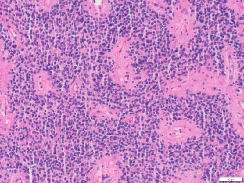
High cell density indicates adverse progression
Posterior fossa ependymoma type A (PF-EPN-A, PFA) often has a poor outcome, with more than half of patients not surviving the disease. They usually occur in young children between the ages of two and five. Swenja Gödicke, like Lara Pohl also a medical student at the University Medical Center Hamburg-Eppendorf (UKE), discovered that the cell density within a tumor differs in these ependymomas. A high cell density is associated with a worse course of the tumor.
The differences between areas of high and low cell density in the tumor tissue sections, which are already visible under the microscope, are reflected at the molecular level. For example, chromosomal abnormalities characteristic of this tumor, which indicate a poor prognosis, were more common in cell-dense areas. Tissue samples from relapses, i.e. recurrences of PF-EPN-A, also showed an increased number of cell-dense areas.
The results of Swenja Gödicke show for the first time how important it is for the assessment of this brain tumor to determine the number of cell-dense areas and to perform molecular analyses of these areas.
"With these three studies, we have made valuable new contributions to the classification of ependymomas. They are also the result of many years of reference pathology work in which we receive ependymomas from all over Germany for evaluation in Hamburg," says Prof. Schüller.
Original publications
Neyazi, S., Yamazawa, E., Hack, K. et al. Transcriptomic and epigenetic dissection of spinal ependymoma (SP-EPN) identifies clinically relevant subtypes enriched for tumors with and without NF2 mutation. Acta Neuropathol 147, 22 (2024). https://doi.org/10.1007/s00401-023-02668-9
Pohl, L.C., Leitheiser, M., Obrecht, D. et al. Molecular characteristics and improved survival prediction in a cohort of 2023 ependymomas. Acta Neuropathol 147, 24 (2024). https://doi.org/10.1007/s00401-023-02674-x
Gödicke, S., Kresbach, C., Ehlert, M. et al. Clinically relevant molecular hallmarks of PFA ependymomas display intratumoral heterogeneity and correlate with tumor morphology. Acta Neuropathol 147, 23 (2024). https://doi.org/10.1007/s00401-023-02682-x
Scientific contact
Prof. Ulrich Schüller
Research Institute Children's Cancer Center Hamburg
Email: schueller@kinderkrebs-forschung.de
Press contact
Research Institute Children's Cancer Center Hamburg
Building N 63
Martinistr. 52
20251 Hamburg
Email: presse@kinderkrebs-forschung.de
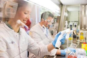
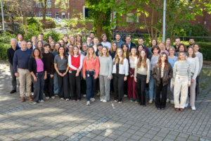
About the Research Institute Children's Cancer Center Hamburg
The Research Institute Children's Cancer Center Hamburg was founded in 2006 by the Fördergemeinschaft Kinderkrebs-Zentrum Hamburg e.V. with donations. At the institute, a multidisciplinary team of around 50 clinically active doctors, scientists, technical assistants, and dedicated employees investigate the molecular mechanisms of cancer development in children in order to develop new approaches for better and more targeted therapies. Donations, sponsorships, and private commitment make the research work possible. The institute is supported by a scientific advisory board and works closely with the University Medical Center Hamburg-Eppendorf (UKE) and the Leibnitz Institute of Virology (LIV). Cooperation with national and international research institutions as well as with clinical patient care at the UKE creates optimal conditions for the successful treatment of children with cancer. Around half of the project costs are covered by competitive third-party funding - including from the German Research Foundation, German Cancer Aid, the Federal Ministry of Education and Research, and the European Union.
About childhood cancer
In Germany, around 2,200 children and adolescents are diagnosed with cancer every year - the most common forms are blood cancers (leukemias), brain tumors, and tumors of the lymphatic tissue. In contrast, carcinomas, which account for more than 90 percent of new cases in adults, are rare. Being diagnosed with cancer is deeply upsetting for families and turns everyday life upside down. Treatment usually lasts for weeks and months. The various forms of therapy are extremely stressful for young patients. Acute side effects are highly likely to occur. Another cause for concern is the increasing number of serious late effects of radiotherapy and chemotherapy for childhood cancer. In general, thanks to successful research in recent decades, great success has been achieved in the fight against childhood cancers. Today, the diseases are curable in many cases: around 80 percent of all those affected survive. This is a great success when you consider that just a few decades ago, these children had hardly any chance of survival. Nevertheless, too many children still die of cancer today or experience a serious loss of quality of life as a result of the disease or the treatment methods.